Faculty Information

Chayan Kanti Nandi
Office:
Personal Information
Professional Experience
Professor, IIT Mandi, (Dec 2020- Till date)
Associate Professor, IIT Mandi (June 2015-Dec 2020)
Assistant Professor, IIT Mandi, (Aug 2010-June 2015)
Postdoctoral Fellow, Princeton University, 2009-2010
Postdoctoral Fellow, Geothe University, 2006-2009
Educational Background
Ph.D., Indian Institute of Technology Kanpur, 2001-2006
M.Sc. (Physical Chemistry) from University of Burdwan, WB, 1998-2000
B. Sc. (Chemistry) from University of Burdwan, WB,1995-1998
Broad Area
Physical Chemistry, Spectroscopy, Microscopy
Specific Areas
Spectroscopy and microscopy of nanomaterials
Designing new fluorescent materials for microscopic imaging
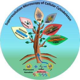
Super resolution imaging of cellular cytoskeleton
Fluorescence lifetime imaging microscopy
Stochastic Optical Reconstruction microscopy
CY-701: Advanced Physical Methods in Chemistry (PhD).
CY 512: Quantum Chemistry (M.Sc)
CY 513: Chemical Thermodynamics (M.Sc)
CY 514: Molecular Reaction Dynamics (M.Sc)
CY 511P: Physical Chemistry lab
IC 101: Basic Chemistry (B. Tech)
IC 101P: Chemistry Lab (B Tech)
IC 201: Design Practicum (B. Tech)
CY 511: Spectroscopy and Molecular Symmetry (M.Sc)
CY670: Advanced fluorescence and microscopy (PhD & M.Sc)
2020 (June): Guest Editor, Frontiers in Chemistry
2019 (July): Chemical Research Society of India (CRSI) bronze medal
2019 (Feb): Best poster award in MRSI-AGM
2018 (June): Visiting Professor, Gottingen University
2017 (sept): Visiting Professor, University of Nottingham.
2017 (Sept): Best Teaching award at IIT Mandi for excellence in Chemistry
2016 (Feb): IIT Mandi Outstanding Faculty for innovative research award
2015 (Sep): DST travel grant awards for conference in Berlin Germany
2015 (Sep): DBT travel grant awards for conference in Berlin Germany
2011 (Dec) : visiting Professor Braunschweig, Germany (by AvH foundation, Germany)
2007-2008: Alexander Von Humboldt Fellow: Geothe University, Frankfurt am Main Germany
2003-2005: Senior Research Fellowship (NET, CSIR, India).
2001-2003: Junior Research Fellowship (NET, CSIR, India).
2001: Graduate Aptitude Test for Engineering India (GATE).
2000: 2nd rank in the M. Sc. Chemistry degree (Burdwan University, WB)
1998: Gold Medal in B. Sc. (Bankura Christian College)
Lifetime members of Materials Research Society of India, 2020
Lifetime members of Electron Microscopy Society of India, 2019
Lifetime Member Chemical Research Society (CRSI) of India 2018
Secretary for "Society for Nanobiotechnology, IIT Mandi (2013)
Chayan Kanti Nandi
Physical Chemistry
Spectroscopy
Microscopy
We work at the interdisciplinary level to fundamentally understand the photophysical properties, especially the photoluminescence origin of florescent nanoprobes for their successful application in single particle level Stochastic Optical Reconstruction Microscopy (STORM), which is an advanced level optical nanoscopic imaging technique. We have developed custom built STORM set up at IIT Mandi to understand the dynamical cellular events under living condition. Currently, we are developing correlative light and electron microscopy (CLEM), which is the combination of both super resolution light microscopy and electron microscopy. CLEM measured the high resolution optical and electron microscopic image from the same spot thus overcoming the individual shortcoming and provide new information on the unknown cellular events. We design carbon based nanoprobes, gold nanoclusters and iron based magneto-fluorescent nanoprobes with a major focus to make them as universal nanoprobes for multimodal single particle level super resolution imaging.
- ACS Materials Letters. 2022, 4, 1565-1573
- Biomaterials Science. 2022, 10, 4525-4537
- Nanoscale 2022, 14, 3568-3578
- Adv. Healthcare Mater. 2022, 2102640
- Chem. Sci. 2021, 12, 3615-3626
- Nat. Commun. 2019, 10 (1), 2391
- Chem. Sci. 2021, 12, 3615-3626
- J. Phys. Chem. C 2021, 125 (3), 1637–1653
- J. Phys. Chem. Lett. 2020, 11 (14), 5741–5748
- Nanoscale 2019, 11 (14), 6561–6565
- Chem. Commun. 2020, 56 (88), 13599–13602
- Chem. Sci. 2017, 9 (1), 175–180
- Nano Lett. 2015, 15 (12), 8300–8305
- 2022
-
- Kaushik, K.; Yadav, A.; Anjum, F.; Mishra, P. M.; Sharma, S.; Rao, C.; Nandi, C. K. Protein Conjugation helped CdTe Quantum Dots for the Specific Labeling and Super-Resolution Imaging of Lysosomes. ChemNanoMat. 2022. https://doi.org/10.1002/cnma.202200235
- Qiu, K.; Yadav, A.; Tian, Z.; Guo, Z.; Shi, D.; Nandi, C. K.; Diao, J. Ultra-Long-Term Super-Resolution Tracking of Lysosomes in Brain Organoids by Near-Infrared Noble Metal Nanoclusters. ACS Materials Letters. 2022, 4, XXX, 1565–1573. https://doi.org/10.1021/acsmaterialslett.2c00436
- Mishra, P. M.; Anjum, F.; Uversky, V. N.; Nandi, C. K. SARS-CoV-2 Spike mutations modify the interaction between virus Spike and human ACE2 receptors. Biochemical and Biophysical Research Communications. 2022, 620, 8-14. https://doi.org/10.1016/j.bbrc.2022.06.064
- Rao, C.; Sharma, S.; Garg, R.; Anjum, F.; Kaushik, K.; Nandi, C. K. Mapping the Time Dependent DNA Fragmentation caused by doxorubicin Loaded on PEGylated Carbogenic Nanodots using Fluorescence Lifetime Imaging and Super-resolution microscopy. Biomaterials Science. 2022, 10, 4525-4537. https://doi.org/10.1039/D2BM00641C
- Yadav, A.; Kaushik, K.; Sharma, S.; Anjum, F.; Nandi, C. K. Near-Infrared-Emitting Ag Nanoclusters as Fluorescent Probes for Super-Resolution Radial Fluctuation Imaging of Lysosomes. ACS Appl. Nano Mater. 2022, 5(7), 9260-9265. https://doi.org/10.1021/acsanm.2c01604
- Singh, S.; Rao, C.; Nandi, C. K.; Mukherjee, T. K. Quantum Dot‐Embedded Hybrid Photocatalytic Nanoreactors for Visible‐light Photocatalysis and Dye Degradation. ACS Appl. Nano Mater. 2022, 5 (5), 7427–7439. https://doi.org/10.1021/acsanm.2c01446
- Yadav, A.; Rao, C.; Kaushik, K.; Anjum, F.; Sharma, S.; Nandi, C. K. Superparamagnetic Iron Oxides Nanoparticles with Large Magnetic Saturation and High Particle Photon Counts for Super Resolution Imaging of Lysosomes. ACS Appl. Nano Mater. 2022, 5 (3), 4018–4027. https://doi.org/10.1021/acsanm.2c00011
- Batra, G.; Sharma, S.; Kaushik, K.; Rao, C.; Kumar, P.; Kumar, K.; Ghosh, S.; Jariwala, D.; Stach, E. A.; Yadav, A.; Nandi, C. K. Structural and Spectroscopic Characterization of Pyrene Derived Carbon Nano Dots: A Single-Particle Level Analysis. Nanoscale 2022, 14, 3568-3578. https://doi.org/10.1039/D1NR07190D
- Gupta, S.; Mishra, D. K.; Khan, M. Z.; Saini, V.; Mehta, D.; Kumar, S.; Yadav, A.; Mitra, M.; Rani, P.; Singh, M.; Nandi, C. K.; Das, P.; Ahuja, V.; Nandicoori, V. K.; Bajaj, A. Development of a Highly Specific, Selective, and Sensitive Fluorescent Probe for Detection of Mycobacteria in Human Tissues. Adv. Healthcare Mater. 2022, 2102640. https://doi.org/10.1002/adhm.202102640
- 2021
-
- Yadav, R.; Yadav, A.; Sharma, S.; Rao, C.; Nandi, C. K. Shedding Light onto the Photoluminesce Origin in Carbon Nanodots Synthesized via Top-down Method. ISRAPS Bulletin 2021, Vol. 33, Issue Number 1. [View]
- Mishra, P. M.; Rao, C.; Sarkar, A.; Yadav, A.; Kaushik, K.; Jaiswal, A.; Nandi, C. K. Super Resolution Microscopy Revealed the Lysosomal Expansion during Epigallocatechin Gallate Mediated Apoptosis. Langmuir 2021, 37 (36), 10818–10826. https://doi.org/10.1021/acs.langmuir.1c01742
- Mishra, P. M.; Nandi, C. K. Structural Decoding of Small Molecular Inhibitor on the Binding of SARS-CoV-2 to ACE2 Receptor. J. Phys. Chem. B 2021, 125 (30), 8395–8405. https://doi.org/10.1021/acs.jpcb.1c03294
- Rao, C.; Patel, S. K.; Prasad, A.; Garg, N.; Nandi, C. K. The Effect of Protein Corona on The Drug Delivery of Carbogenic Nanodots and their Mapping by Fluorescence Lifetime Imaging Microscopy. ACS Appl. Bio Mater. 2021, 4 (7), 5776–5785. https://doi.org/10.1021/acsabm.1c00526
- Wang, F.; Yang, X.; Zhan, Q.; Nandi, C. K. Recent Advances in Fluorescent Probes for Super-Resolution Microscopy. Front. Chem., 11 June 2021. https://doi.org/10.3389/fchem.2021.698531
- Batra, G.; Sharma, S.; Kaushik, K.; Rao, C.; Kumar, P.; Kumar, K.; Ghosh, S.; Jariwala, D.; Stach, E. A.; Yadav, A.; Nandi, C. K. Structural and Spectroscopic Characterization of Pyrene Derived Carbon Nano Dots: A Single Particle Level Analysis. ChemRxiv. Cambridge: Cambridge Open Engage; 2021 (Pre-Print). https://doi.org/10.33774/chemrxiv-2021-8gl92-v2
- Soni, N.; Singh, S.; Sharma, S.; Batra, G.; Kaushik, K.; Rao, C.; Verma, N. C.; Mondal, B.; Yadav, A.; Nandi, C. K. Absorption and Emission of Light in Red Emissive Carbon Nanodots. Chem. Sci. 2021, 12, 3615-3626. https://doi.org/10.1039/d0sc05879c
- Verma, N. C.; Yadav, A.; Rao, C.; Mishra, P. M.; Nandi, C. K. Emergence of Carbon Nanodots as a Probe for Super-Resolution Microscopy. J. Phys. Chem. C 2021, 125 (3), 1637–1653. https://doi.org/10.1021/acs.jpcc.0c09695
- 2020
-
- Yadav, A.; Rao, C.; Nandi, C. K. Fluorescent Probes for Super-Resolution Microscopy of Lysosomes. ACS Omega 2020, 5 (42), 26967–26977. https://doi.org/10.1021/acsomega.0c04018
- Tiwari, A.; Verma, N. C.; Turkkan, S.; Debnath, A.; Singh, A.; Draeger, G.; Nandi, C. K.; Randhawa, J. K. Graphitic Carbon Coated Magnetite Nanoparticles for Dual Mode Imaging and Hyperthermia. ACS Appl. Nano Mater. 2020, 3 (1), 896–904. https://doi.org/10.1021/acsanm.9b02501
- Kumar, P.; Thakar, K.; Verma, N. C.; Biswas, J.; Maeda, T.; Roy, A.; Kaneko, K.; Nandi, C. K.; Lodha, S.; Balakrishnan, V. Polymorphic In-Plane Heterostructures of Monolayer WS2 for Light-Triggered Field-Effect Transistors. ACS Appl. Nano Mater. 2020, 3 (4), 3750–3759. https://doi.org/10.1021/acsanm.0c00027
- Mani Mishra, P.; Uversky, V. N.; Nandi, C. K. Serum Albumin-Mediated Strategy for the Effective Targeting of SARS-CoV-2. Med. Hypotheses 2020, 140, 109790. https://doi.org/10.1016/j.mehy.2020.109790
- Yadav, A.; Rao, C.; Verma, N. C.; Mishra, P. M.; Nandi, C. K. Magnetofluorescent Nanoprobe for Multimodal and Multicolor Bioimaging. Mol. Imaging 2020, 19, 153601212096947. https://doi.org/10.1177/1536012120969477
- Rao, C.; Mishra, P. M.; Yadav, A.; Nandi, C. K. Cancer Cell Membrane Technology for Cancer Therapy. ChemNanoMat 2020, 6 (12), 1712–1729. https://doi.org/10.1002/cnma.202000482
- Yadav, A.; Verma, N. C.; Rao, C.; Mishra, P. M.; Jaiswal, A.; Nandi, C. K. Bovine Serum Albumin-Conjugated Red Emissive Gold Nanocluster as a Fluorescent Nanoprobe for Super-Resolution Microscopy. J. Phys. Chem. Lett. 2020, 11 (14), 5741–5748. https://doi.org/10.1021/acs.jpclett.0c01354
- Rao, C.; Yadav, A.; Kaur, R.; Prasad, A.; Nandi, C. K. Direct Visualization of the Protein Corona Using Carbon Nanodots as a Specific Contrasting Agent. Chem. Commun. 2020, 56 (88), 13599–13602. https://doi.org/10.1039/d0cc06333a
- 2019
-
- Rao, C.; Singh, A.; Verma, N. C.; Garg, N.; Nandi, C. K. One Pot Synthesis of Amphiphilic Carbogenic Fluorescent Nanodots for Bioimaging. ChemNanoMat 2019, 5 (4), 417–421. https://doi.org/10.1002/cnma.201800663
- Butkevich, E.; Verma, N. C.; Oleksiievets, N.; Gregor, I.; Schmidt, C. F.; Enderlein, J.; Nandi, C. K.; Chizhik, A. I. Carbon Dots for Studying Muscle Architecture. ACS Appl. Nano Mater. 2019, 2 (12), 7466–7472. https://doi.org/10.1021/acsanm.9b01815
- Rao, C.; Verma, N. C.; Nandi, C. K. Unveiling the Hydrogen Bonding Network of Intracellular Water by Fluorescence Lifetime Imaging Microscopy. J. Phys. Chem. C 2019, 123 (4), 2673–2677. https://doi.org/10.1021/acs.jpcc.8b12439
- Tiwari, A.; Verma, N. C.; Randhawa, J. K.; Nandi, C. K. Real-Time Observation of Magnetic Field-Induced Fluorescence Engineering in SPIONs. J. Phys. Chem. C 2019, 123 (45), 27759–27764. https://doi.org/10.1021/acs.jpcc.9b07261
- Verma, N. C.; Rao, C.; Singh, A.; Garg, N.; Nandi, C. K. Dual Responsive Specifically Labelled Carbogenic Fluorescent Nanodots for Super Resolution and Electron Microscopy. Nanoscale 2019, 11 (14), 6561–6565. https://doi.org/10.1039/c9nr00457b
- Verma, N. C.; Yadav, A.; Nandi, C. K. Paving the Path to the Future of Carbogenic Nanodots. Nat. Commun. 2019, 10 (1), 2391. https://doi.org/10.1038/s41467-019-10394-8
- 2018
-
- Khan, S.; Verma, N. C.; Chethana; Nandi, C. K. Carbon Dots for Single-Molecule Imaging of the Nucleolus. ACS Appl. Nano Mater. 2018, 1 (2), 483–487. https://doi.org/10.1021/acsanm.7b00175
- Kumar, P.; Verma, N. C.; Goyal, N.; Biswas, J.; Lodha, S.; Nandi, C. K.; Balakrishnan, V. Phase Engineering of Seamless Heterophase Homojunctions with Co-Existing 3R and 2H Phases in WS2 Monolayers. Nanoscale 2018, 10 (7), 3320–3330. https://doi.org/10.1039/c7nr08303c
- Verma, N. C.; Rao, C.; Nandi, C. K. Nitrogen-Doped Biocompatible Carbon Dot as a Fluorescent Probe for STORM Nanoscopy. J. Phys. Chem. C 2018, 122 (8), 4704–4709. https://doi.org/10.1021/acs.jpcc.7b12773
- Dwivedi, C.; Chaudhary, A.; Srinivasan, S.; Nandi, C. K. Polymer Stabilized Bimetallic Alloy Nanoparticles: Synthesis and Catalytic Application. Colloids Interface Sci. Commun. 2018, 24, 62–67. https://doi.org/10.1016/j.colcom.2018.04.001
- Nandi, C. K. Mechanistic Insight into the Carbon Dots: Protonation Induced Photoluminescence. J Mater. Sci Eng 2018, 7 (3), 448. https://doi.org/10.4172/2169-0022.1000448
- Tiwari, A.; Verma, N. C.; Singh, A.; Nandi, C. K.; Randhawa, J. K. Carbon Coated Core-Shell Multifunctional Fluorescent SPIONs. Nanoscale 2018, 10 (22), 10389–10394. https://doi.org/10.1039/c8nr01941j
- Khan, S.; Jain, S.; Nandi, C. K. Towards Understanding Citric Acid Derived High Quantum Yield Molecular Fluorophores: From Carbon Dots to Spherical Organic Nanocrystals. J Mater. Sci Eng 2018, 7 (5), 490. https://doi.org/10.4172/2169-0022.1000490